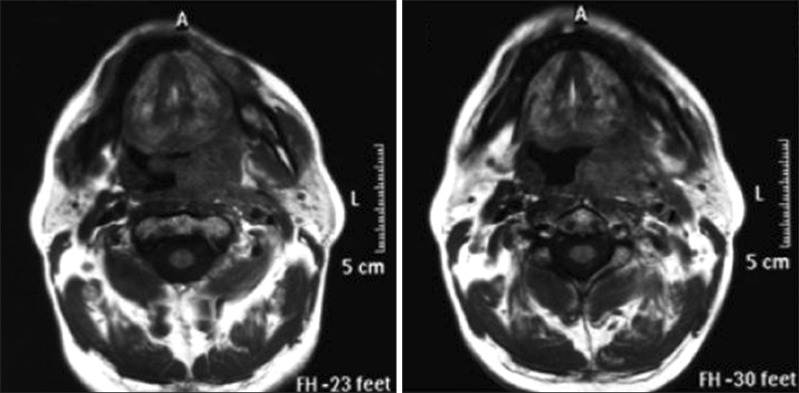
Fig. 1.
The patient's throat magnetic resonance imaging scan showing a heterogeneously dense mass, sized approximately 65×29 mm at its broadest point, with its borders not clearly identified; therefore, the mass caused distinct asymmetry in the hypopharynx and oropharynx, displaced the uvula to the right side, and continued to the epiglottis level.

