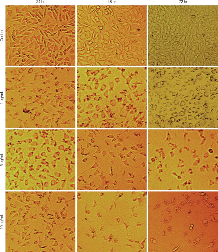
Figure 1.
Morphological alterations of MCF-7 cells after the exposure to different concentrations of cytotoxin-II, observed by a normal inverted light microscopy. Detachment of cells from the dish, cell rounding, cytoplasmic blebbing, chromatin condensation and irregularity in shape are observable. Dose and time-dependent decrease in cell counts was seen (×100).

