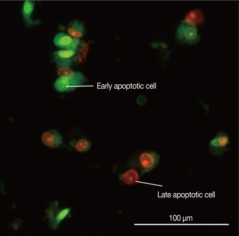
Figure 3.
Picture of MCF-7 cells exposed to cytotoxin-II for 24 hours and stained with acridine orange/ethidium bromide. Early apoptotic cells are shown as bright green chromatin that is highly condensed or fragmented. Late apoptotic cells have bright orange chromatin that is highly condensed or fragmented.

