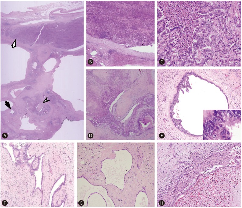
Figure 3.
Microscopic findings (A-H). (A) Scanning power photomicrograph demonstrating an overview of the boxed area of Figure 2. Cystically dilated bile ducts are seen, and a large bile duct (white arrow) is plugged with a cholangiocarcinoma, corresponding to the bright yellow solid lesion on gross examination. The neoplastic lesion is a gland-forming moderately differentiated adenocarcinoma with intraductal growth (B, C). Another bile duct (white arrowhead) in (A) demonstrates features of ductal plate malformation and involvement by cholangiocarcinoma. This area is seen in detail in (D); the fibrovascular intraluminal projection is evident. A smaller peripheral duct (black arrow) in (A) is lined by biliary intraepithelial neoplasia (BilIN) 3, and is seen in detail in (E). A micropapillary architecture is seen in addition to the high-grade nuclear atypia (inset). Features of ductal plate malformation were seen in other large and small bile ducts (F, G). (H) demonstrates a smaller bile duct that was affected by ascending cholangitis: the mucosa is ulcerated and the lumen is filled with inflammatory exudate. [Hematoxylin-eosin, original magnification ×1.25 (A), ×40 (B, D), ×100 (E, F, H), ×200 (G), ×400 (C, inset of E)].

