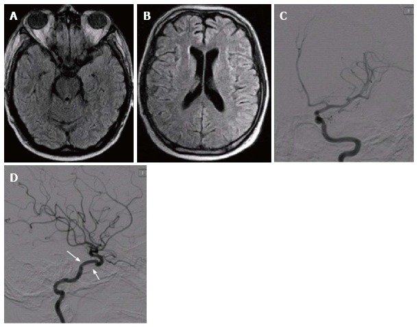
Figure 2.

Three months follow up evaluation shows a normal brain magnetic resonance imaging (FLAIR A and B); left internal carotid angiogram (C: AP view, D: Lateral view) shows a normal flow through the Jostent Graftmaster in the cavernous segment (white arrows).
