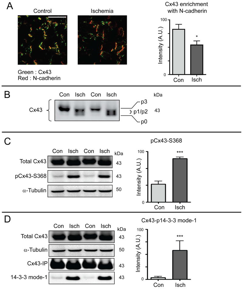
Figure 4. Acute cardiac ischemia induces hyper-phosphorylation of Cx43 at serines 368 and 373.
Hearts from 8-week-old C57BL/6 mice were maintained using a Langendorff perfusion apparatus for 15 minutes, followed either by 30 minutes of normal perfusion (control) or no-flow ischemia (n = 3 per condition). A) Confocal immunofluorescence of Cx43 (green) and N-cadherin (red) in cryosections from snap-frozen hearts. Quantification of Cx43/N-cadherin colocalization in graph on right. Original magnification: X60. Scale bar = 50 μm. B) Western blot of lysates from control (con) and ischemic (isch) hearts probed for Cx43 to visualize phosphospecific forms p0 – p3. C) Western blot of lysates from control and ischemic hearts probed for total Cx43 (top panel), pCx43Ser368 (middle panel) and α-tubulin (bottom panel). Quantification of pCx43Ser368 relative to total Cx43 in graph on right. D) Cx43 immunoprecipitations from control and ischemic hearts. Immunoprecipitation Western blot was probed for Cx43 and phosphorylated 14-3-3 mode-1 motif. Quantification of phosphorylated 14-3-3 mode-1 motif in graph on right.

