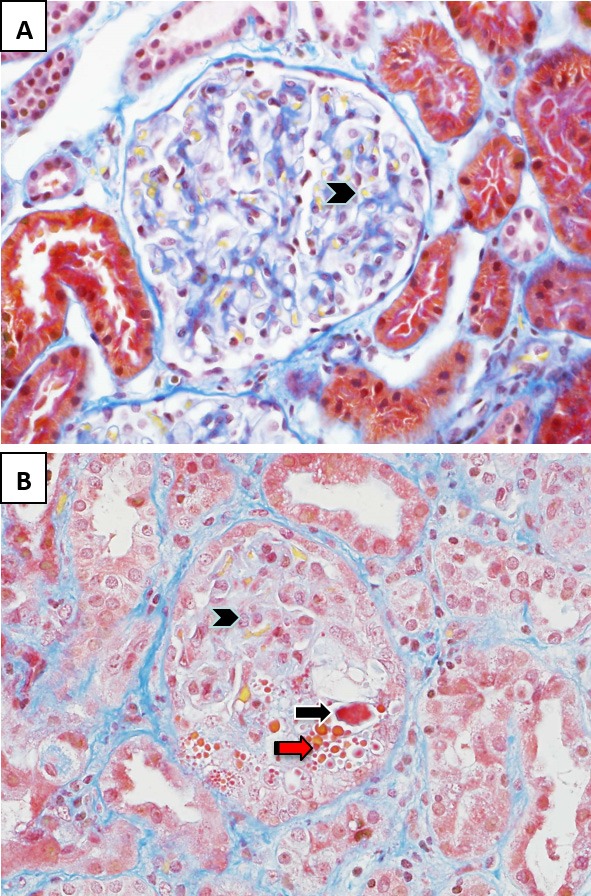
Figure 1. Glomerular pathology in hemolytic uremic syndrome.

(A) In the normal glomerulus, patent capillary lumina containing erythrocytes stain yellow (arrowhead). (B) In hemolytic uremic syndrome, the glomerular capillary loops contain fibrin thrombi and microthrombi that stain bright red (black arrow). There is endothelial cell swelling with obliteration of some of the capillary lumina (arrowhead) and red cell fragmentation (red arrow). Stain: Martius scarlet blue trichrome; magnification: 400×.
