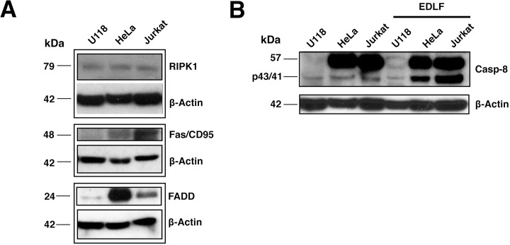
Figure 6. Relative protein levels of RIPK1, Fas/CD95, FADD and procaspase-8 in U118, HeLa and Jurkat cells.
(A) U118, HeLa and Jurkat cell lines were analyzed by immunoblotting using specific antibodies against RIPK1, Fas/CD95 and FADD. Immunoblotting for β-actin was used as an internal control for equal protein loading in each lane. (B) U118, HeLa and Jurkat cells untread and treated with 10 μM edelfosine (EDLF) for 24 h were analyzed by immunoblotting using a specific antibody that recognizes full-length 57-kDa procaspase-8 and p43/41 cleaved active caspase-8 fragments. The molecular weight of each immunodetected band is indicated. Data shown are representative of three experiments.

