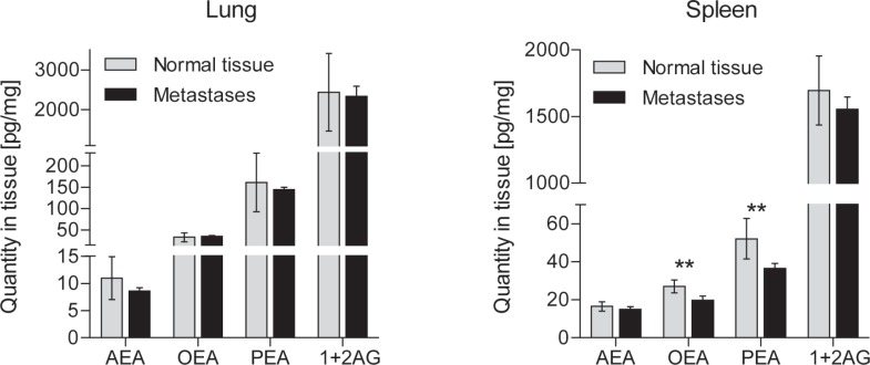
Figure 4. Tissue concentrations (mean ± SD) of endocannabinoids in metastatic mouse B16 mouse melanoma.
Cells were injected via the tail vein and metastases were identified on the basis of the melanin production. They developed in lung, spleen and lymph nodes. Metastases and corresponding control tissue samples were dissected at 4 weeks (n = 10 mice for plasma, 5-8 for tissue). Asterisks indicate significant differences versus baseline (plasma) or metastasis versus non-cancerous corresponding tissues (t-tests after significant ANOVA, p < 0.05).

