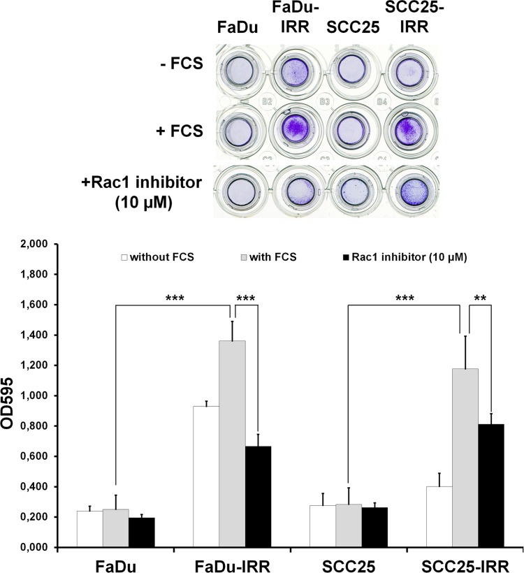
Figure 7. Effects of Rac1 inhibitor on HNSCC cell migration.
Differences in migration of parental and IRR HNSCC cells, and Rac1 inhibitor-induced repression of cell migration, were determined using a QCMTM 24-well colorimetric cell migration assay (Merck Millipore, Darmstadt, Germany), following the manufacturer's instructions. HNSCC cells harvested in the appropriate serum-free quenching medium were placed in the upper insert with an 8-μm pore size polycarbonate membrane. The lower chamber contained culture medium with chemoattractant (10% FCS). Plates were incubated for 24 hours at 37°C in a 5% CO2 humidified atmosphere. HNSCC cells that migrated through the membrane were stained and then subsequently extracted using extraction buffer. The optical densities of dye extracts were read at 560 nm using a microplate reader (Bio-Rad Microplate Reader 680, Bio-Rad Laboratories GmbH, Munich, Germany). **p < 0.01; ***p < 0.001 [9].

