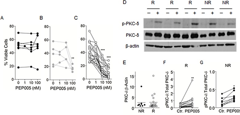
Figure 3. PEP005 treatment leads to the activation of PKC-δ in both responsive and non-responsive patient samples.
Primary AML samples were grouped into non-responders (A), low-responders (B) or responders (C) based on their apoptotic response to PEP005 treatment. Apoptosis was measured using Annexin V/PI staining. D. Isolated AML blasts were cultured in medium alone (−) or 10 nM PEP005 (+) for 30 minutes. SDS-PAGE and western blotting were then used to measure phospho-PKC-δ and PKC-δ expression, with β-actin used as a loading control. Samples that did not respond to PEP005 treatment in the apoptosis experiments were termed non-responders (NR), and those that did respond were termed responders (R). E–G. Densitometric analysis was used to determine the ratio of total PKC-δ to β-actin (E) and phosphorylated to total PKC-d in both R (F) and NR (G) samples. * indicates p<0.05, ** indicates p<0.01.

