
Figure 8.
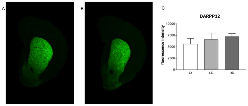
Quantitative infrared immunofluorescence analysis of striatal DARPP32 levels. Coronal slices from rat striatum (STR) were probed for antibodies against DARPP32. Secondary antibodies were tagged with an infrared fluorescent marker. Depicted are representative scans showing fluorescence intensity in STR slices from control animals (A) and Mn treated animals (B). A minimum of three slices were quantified from each animal and the resulting values were averaged. Group values were averaged and are shown (C). No difference was observed between control and either low or high dose groups. Mn treatment did not affect STR DARPP32 signal intensity. ANOVA with Tukey’s post hoc test for multiple comparisons. Data represent mean ± SD (n = 4–5).