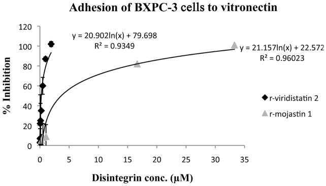
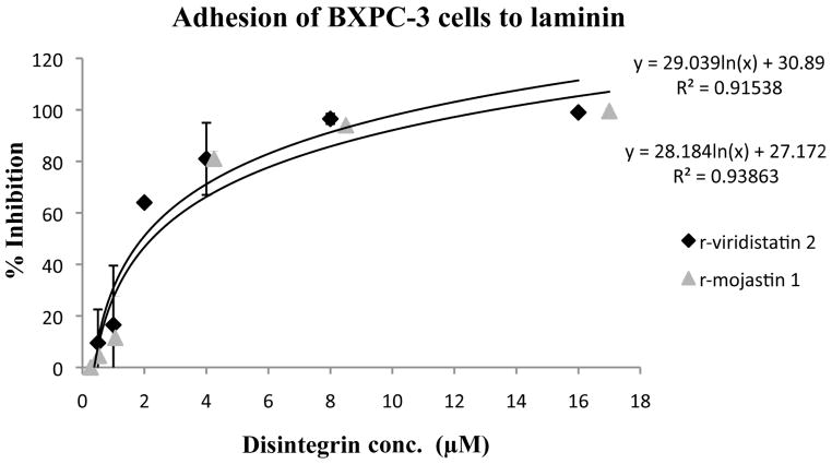
Figure 1.

Inhibition of BXPC-3 cells adhesion to laminin (A) and vitronectin (B) in vitro by r-viridistatin 2 and r-mojastin 1. BXPC-3 cells (5×10⁵ cells/mL, 0.2 mL) were treated with different concentrations of r-disintegrins for 1 h at 37 °C, and then added to a 96 well-plate coated with extracellular matrix protein (10 μg/mL). After removal of non-bind cells, the remaining cells were quantified using 3-[4,5-dimethylthiazol-2-yl] 2,5-diphenltetrazolium bromide (MTT) and the IC50 values calculated as described in methods section. Graph equation: y: 50% proliferation; x: disintegrin concentration; R2: square of coefficient of correlation.

