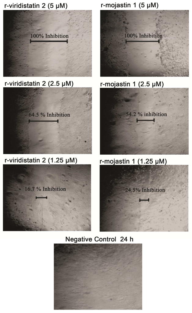
Figure 3.
Inhibition cell migration of BXPC-3 cells. A confluent monolayer of cells was maintained in medium, and a line was scraped through the monolayer of cells with a plastic, sterile pipette tip. The cultures were allowed to migrate for 24 h at 37°C in the presence or absence of recombinant disintegrins at 1.25, 2.5, and 5 μM. The extent of wound closure was quantified by multiple measurements of the width of the scrape space for each cell line.

