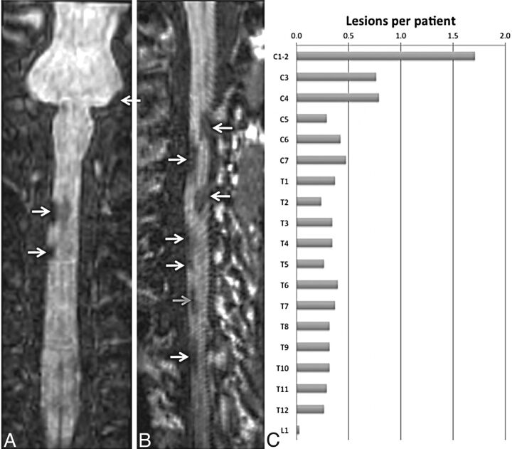
Fig 4.
Coronal (A) and curved-reformatted (B) images from the T1-weighted MPRAGE sequence in the cervical cord of 2 patients, showing lesions mainly on the surface of the cord (white arrows) and a lesion that appears to be completely within the cord (gray arrow). C, Incidence of lesions per vertebral body segment derived from all scans acquired in the MS population studied herein (Table, 39 patients with MS).

