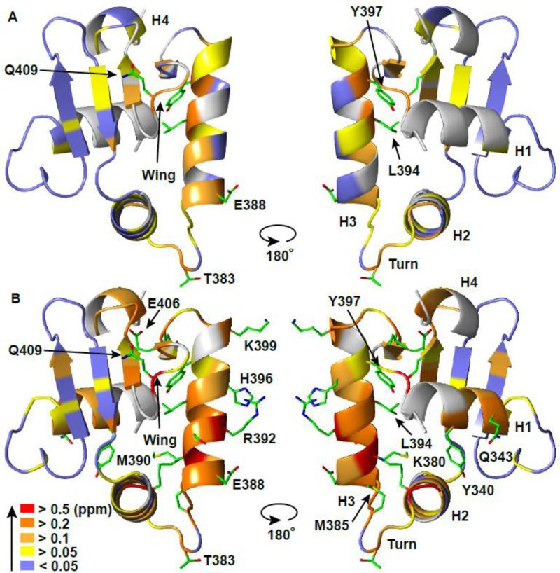
Fig. 5. ETV6 ETS domain binds specific and non-specific DNA via the same canonical interface.
The amide chemical shift perturbations (CSP's, Δδ = {(ΔδH2 + (0.154ΔδN)2}1/2) for (A) ETV6D446–DNAnonsp and (B) ETV6D446–DNAsp with respect to unbound ETV6R426 are mapped onto the crystal structure of ETV6R426-DNAsp-cryst (DNA not shown). Residues (backbone cartoon) are color-coded in the indicated CSP ranges. Prolines and unassigned residues are in grey. Side chains are shown for residues in (A) with CSP > 0.2 ppm and in (B) with CSP > 0.4 ppm. See Figure 1 and Supplemental Figures S1 and S4 for the original data.

