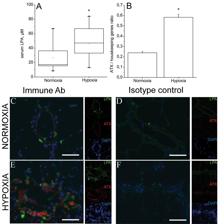
Figure 2.
A, Lysophosphatidic acid (LPA) content in serum from rats kept in ambient air and rats maintained in normobaric hypoxia (10% O2) for 3 weeks. Data obtained from normoxia group of animals deviated significantly from normal distribution (Kolmogorov-Smirnov test). Accordingly, the values from both groups were log transformed before statistical analysis. After detransformation, mean values were 21.6 (11.0–42.3) μM for the normoxia group and 40.9 (23.4–71.7) μM for the hypoxia group (P = 0.037 on lognormal data, nonpaired t test). B, Expression of lysophospholipase (autotaxin [ATX]) in lungs of normoxic and hypoxic rats. Data represent mean + SD of ATX messenger RNA (mRNA) expression normalized to geometric mean of mRNA expression of 2 housekeeping genes (GAPDH and HPRT). C–F, Immunohistochemical analysis of LPA and ATX expression in lungs of normoxic (C, D) and hypoxic (E, F) rats. Green, red, and blue channels are shown separately as thumbnails, and the large images show merged channels in the presence of immune antibodies (C, E) and isotype control antibody (D, F). DAPI: 4′,6-diamidino-2-phenylindole. Scale bars: 25 μm.

