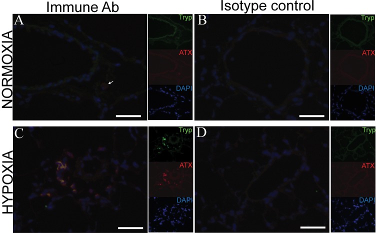
Figure 3.
Immunohystochemical analysis of mast cell tryptase (Tryp) and autotaxin (ATX) expression in lungs of normoxic (A, B) and hypoxic (C, D) rats. Green, red, and blue channels are shown separately as thumbnails, and the large images show merged channels in the presence of immune antibodies (A, C) and isotype control antibody (B, D). The arrow in A points to a single weakly ATX-positive cell in the lung of a normoxic rat. DAPI: 4′,6-diamidino-2-phenylindole. Scale bars: 25 μm.

