Figure 3.
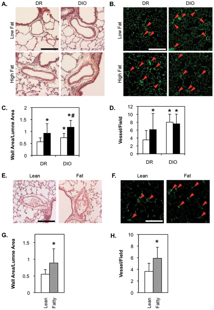
PA remodeling and muscularization of distal arterioles in diet-resistant/diet-induced obesity (DR/DIO) and Zucker lean/fatty rat models. A–D, Eight-week-old male DR and DIO rats were fed either a low-fat (white bars in C, D) or a high-fat (black bars in C, D) diet ad lib. for 5 months. E–H, Eight-week-old male Zucker lean and fatty rats were fed standard chow ad lib. for 5 months. A, E, Representative bright-field micrographs of hematoxylin-and-eosin-stained lung sections show medial thickening in high-fat-fed DR and low- and high-fat-fed DIO rats (A) and in Zucker fatty rats (E). Scale bars = 100 μm. C, G, Remodeling was confirmed by morphometric analysis of 10 pulmonary arteries per animal. B, D, Representative fluorescence micrographs of lung parenchyma stained with an anti-rat smooth muscle (SM)-actin antibody, followed by an Alexa Fluor 488–conjugated secondary antibody. Arrows indicate scored actin-positive microvessels. Scale bars = 50 μm. D, H, Morphometric analysis of the number of actin-positive vessels per field in 10 fields per animal. Values in C, D, G, and H are averages from 3 experiments with 6 animals per group.

