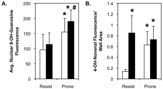
Figure 7.

Morphometric analysis confirms oxidant damage in the pulmonary artery (PA) wall of diet-induced-obesity-resistant (Resist) and diet-induced-obesity-prone rats. Nuclear 8-OH-guanosine (A) and total medial 4-OH-nonenal (B) levels were quantified by morphometric analysis of fluorescent images like those in Figure 6. The relative 8-OH-guanosine fluorescence was measured in 6–8 individual nuclei (identified by 4,6-diamidino-2-phenylindole [DAPI] fluorescence) in 10 PAs in lung sections from 6 rats in each group from 2 separate experiments. Relative 4-OH-nonenal fluorescence was measured over the entire region defined by smooth muscle (SM)-actin staining in 10 PAs in lung sections from 6 rats in each group from 2 separate experiments. Fluorescence values represent single-channel luminance. White bars: rats fed low-fat diet; black bars: rats fed high-fat diet.
