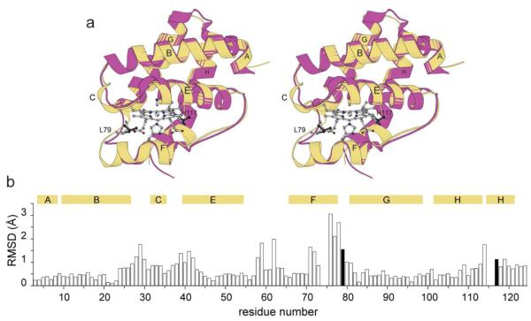
Fig. 2.
Structural comparison of cyanomet C. eugametos L1637 CtrHb (PDB ID: 1DLY) with cyanomet Synechocystis sp. PCC 6803 GlbN-A (PDB ID: 1S69). (a) Stereo view of the superimposition of the two structures. Synechocystis GlbN-A is in yellow with L79 and H117 shown in light gray sticks. Dark gray sticks mark L75 and T111 in CtrHb. (b) Cα rmsd for the superimposition shown in (a). The numbering is that of GlbN. The black vertical bars indicate the position of L79 and H117. The secondary structure of GlbN is indicated with horizontal bars above the rmsd values.

