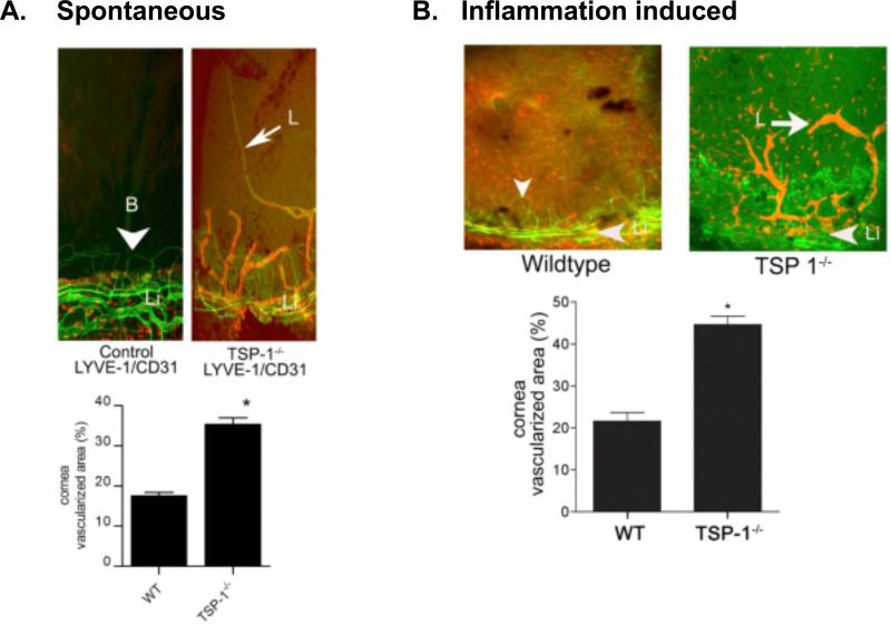
Figure 2. Increased lymphangiogenesis in TSP-1 deficient cornea.
Corneal flat mounts from WT and TSP-1 deficient mice stained with fluorescence conjugated anti-CD31 (green) and anti-LYVE-1 (red) under spontaneous (A) and suture-induced inflammatory conditions (B). Morphometric analysis of vascularized areas with LYVE-1+++ /CD31+ lymphatic vessels is presented below each set of staining. (* p < 0.05). From Cursiefen et al. J.Exp.Med. 2011, 208(5):1083

