Fig. 1.
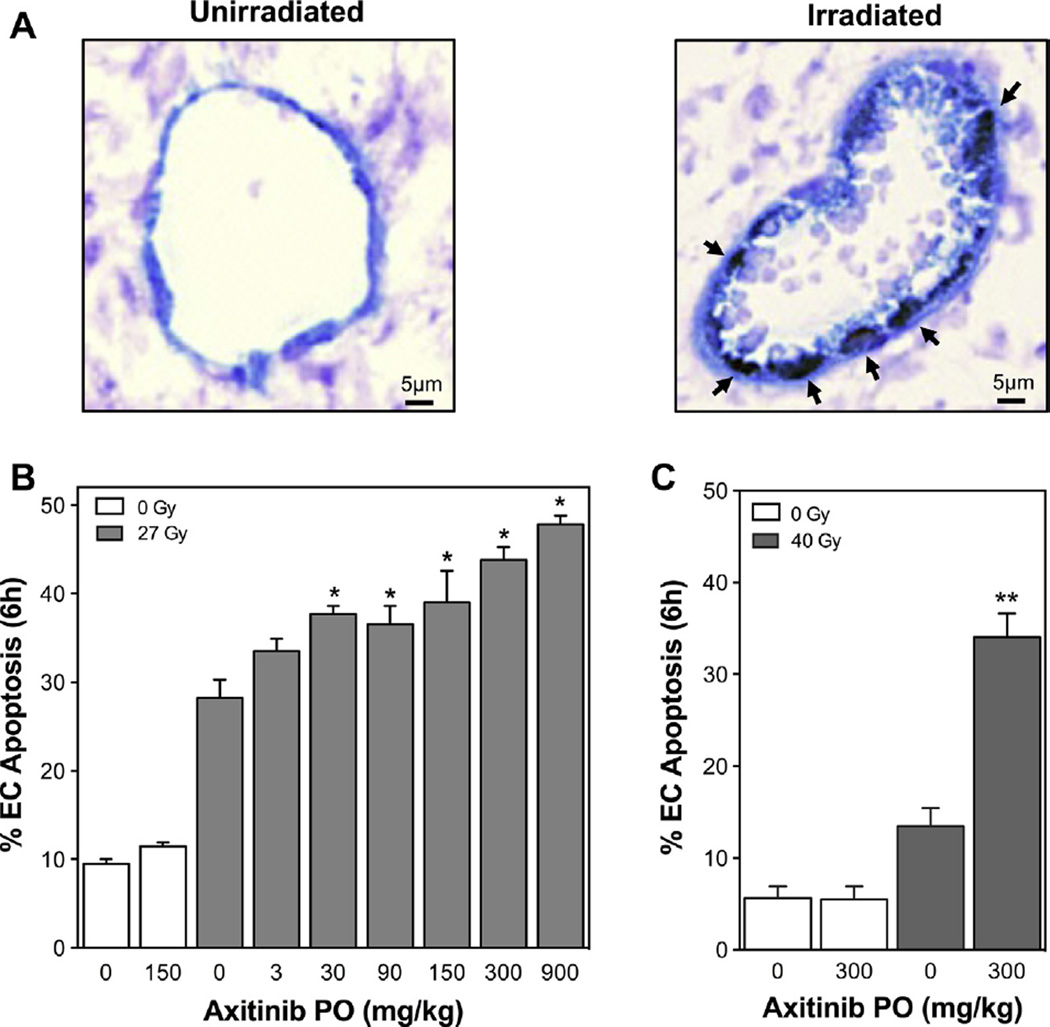
Axitinib increases endothelial cell apoptosis after SDRT in vivo. Axitinib was administered to Sv129/BL6 mice bearing MCA/129 sarcoma or B16F1 melanoma flank tumors (100–150 mm3) and after 1 h tumors were treated with SDRT. Tumors were double-stained using TUNEL labeling and MECA-32 immunohistochemistry to identify apoptotic endothelial cells 6 h after SDRT. (A) Representative 5-µm sections of MCA/129 tumors untreated or at 6 h after 27 Gy SDRT plus axitinib. Apoptotic endothelium exhibits a brown TUNEL-positive nuclear signal surrounded by a dark blue plasma membrane signal for MECA-32 (indicated by arrows, magnification 400×). (B) Axitinib given to mice bearing MCA/129 tumors at 1 h preceding 27 Gy dose-dependently increases SDRT-induced endothelial cell apoptosis. (C) Axitinib administered 1 h preceding 40 Gy to mice bearing B16F1 tumors increases endothelial cell apoptosis. Data (mean ± SEM) are collated from 2 to 3 mice per dose with 1000–2000 endothelial cells evaluated. *p < 0.001 compared to 27 Gy alone. **p < 0.001 compared to 40 Gy alone.

