Fig. 1.
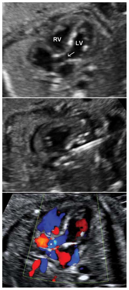
Top panel: Aortic stenosis and evolving hypoplastic left heart syndrome at 25 weeks gestational age. Left ventricular size is preserved in midgestation. Areas of left ventricular echo brightness consistent with endocardial fibroelastosis and myocardial scarring are evident. The thickened, doming, and stenotic aortic valve is visible. Middle panel: The cannula is directed into the left ventricular outflow tract. A guidewire is advanced into the proximal ascending aorta, and a coronary angioplasty balloon is inflated across the aortic valve. Bottom panel: Still-frame color Doppler image demonstrating a broad jet of anterograde flow (asterisk) across the left ventricular outflow tract and aortic valve. LV, left ventricle; RV, right ventricle; arrow, stenotic aortic valve.

