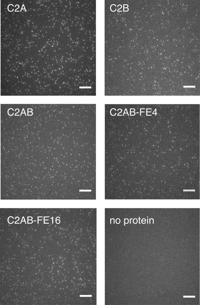
Figure 3.
Images of protein diffusion. All images are taken from movies acquired with 50 ms exposure and illustrate random, uniform distributions of particles with negligible background contamination. The bottom right panel shows the same bilayer as that in the bottom left panel but prior to the addition of protein. Scale bars: 10 μm. Representative movies are available in the Supporting Information.

