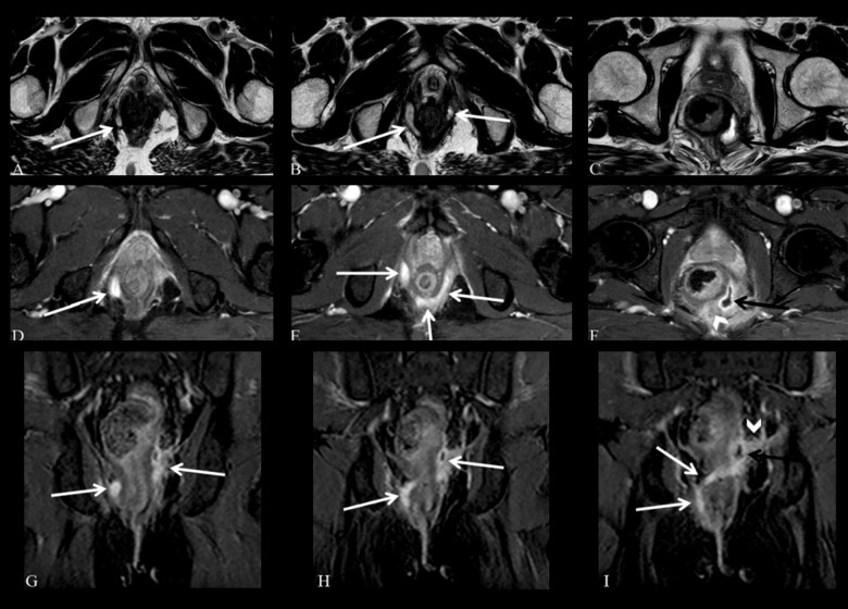
Figure 9.
Grade 5 perianal fistula; supralevator and translevator disease. Axial T2-weighted (A–C) and fat-suppressed T2-weighted (D–F), coronal contrast-enhanced fat-suppressed T1-weighted (G–I) MR images show a right translevator fistula (arrows) crossing the ischiorectal fossa which enters the intersphicteric space posterior to the anal canal and continues with a left supralevator abscess (black arrows) with inflammatory changes surrounding the rectum (arrowheads).

