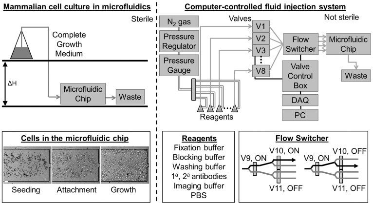
Figure 1. Design of microfluidic platform.
Left, top: The cell culture is performed under sterile conditions. The flow is established using gravity-driven flow and the flow rate is adjusted by changing the value of ΔH. Left bottom: Cells are introduced into the microfluidic chip (seeding), allowed to attach (attachment), and then maintained under steady, continuous perfusion (growth). Right, top: During the data acquisition phase, the chip is removed from the sterile environment, placed on the microscope stage, and connected to a computer-controlled fluid delivery system. The number of reagents can be adjusted by adding or removing valves. Pressurized air is coupled to reagents which are routed through the system using solenoid pinch valves. Right, bottom: Each reagent can be delivered to each channel in the microfluidic chip through a binary multiplexer tree. By adjusting the configuration of the valves, fluid can be routed to different channels (shown are two examples). A total of n-1 three-way solenoid valves are needed to split the incoming fluid from one channel to n channels.

