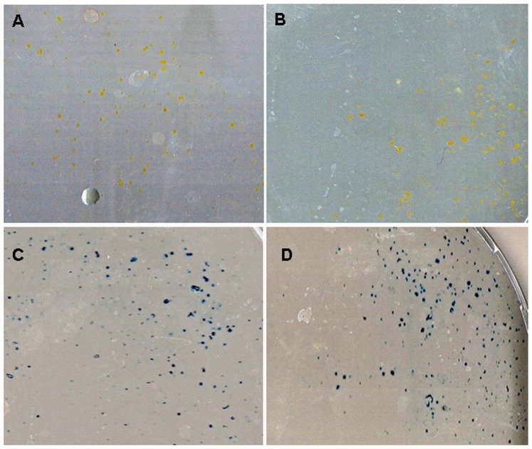
Figure 6. Collagen and methylene blue staining of MuMac-E8 cells.
Used cell number was 1×10⁴. (A) The cultivation was carried out in osteogenic differentiation medium and in (B) normal medium. Isolated yellow-colored cells are visible in both cases. (C) The cultivation was carried out in osteogenic differentiation medium and in (D) normal medium. Isolated blue-colored cells are visible in both cases. There was no colony formation.

