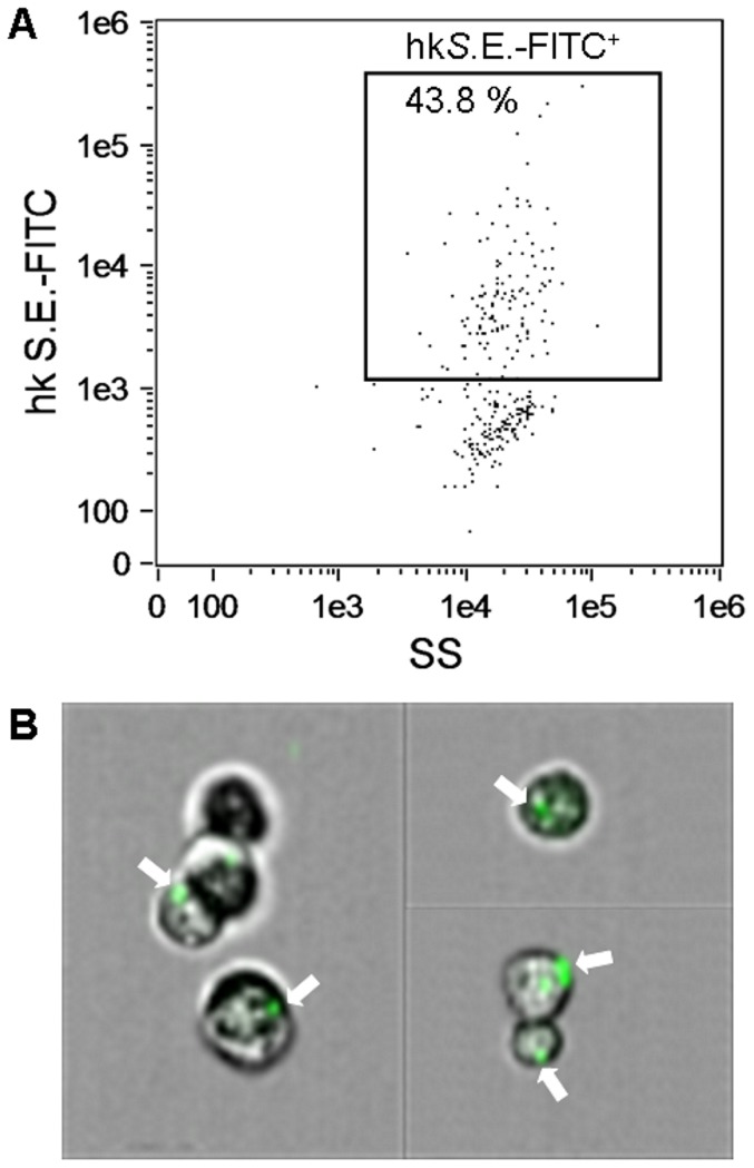
Figure 9. Phagocytic activity of MuMac-E8 cells.
For measurement of phagocytic potential, MuMac-E8 cells were harvested and 1×10⁶ cells were incubated for 2 h with 2×10⁷ FITC-labeled heat-killed salmonellae. Afterwards cells were washed 4 times with HBSS and the uptake of bacteria was assessed by imaging flow cytometry (Amnis FlowSight system, Merck Millipore). In this representative experiment 44% of the cells revealed a positive signal (A). By parallel imaging it could be confirmed that the positive fluorescence signal coincided with phagocytosis of salmonellae. White arrows show cells with internalized FITC-labeled bacteria (B).

