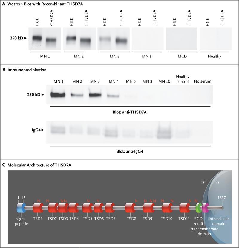
Figure 2. Identification of the Target Antigen.
Panel A shows reactivity of serum samples with human glomerular extracts (HGE) and recombinant thrombospondin type-1 domain-containing 7A (rTHSD7A). Only samples from patients with membranous nephropathy (MN) that previously recognized the 250-kD protein present in HGE also recognized rTHSD7A (MN 1, MN 2, and MN 3). Panel B shows results of immunoprecipitation experiments. All serum samples from patients with membranous nephropathy that previously recognized rTHSD7A precipitated the antigen from HGE. No immunoprecipitation occurred with serum from healthy controls or with no serum (i.e., with water substituted for serum in the experiment). IgG4 was efficiently immunoprecipitated in all conditions (lower panel). Panel C shows the molecular architecture of THSD7A, with a large extracellular region comprising 11 thrombospondin type-1 domains (TSDs), 14 glycosylation sites (N), and 1 predicted arginine–glycine–aspartic acid (RGD) motif. The scheme was built according to the UniProt accession number Q9UPZ6 for THSD7A and ScanProsite tool from ExPASy.

