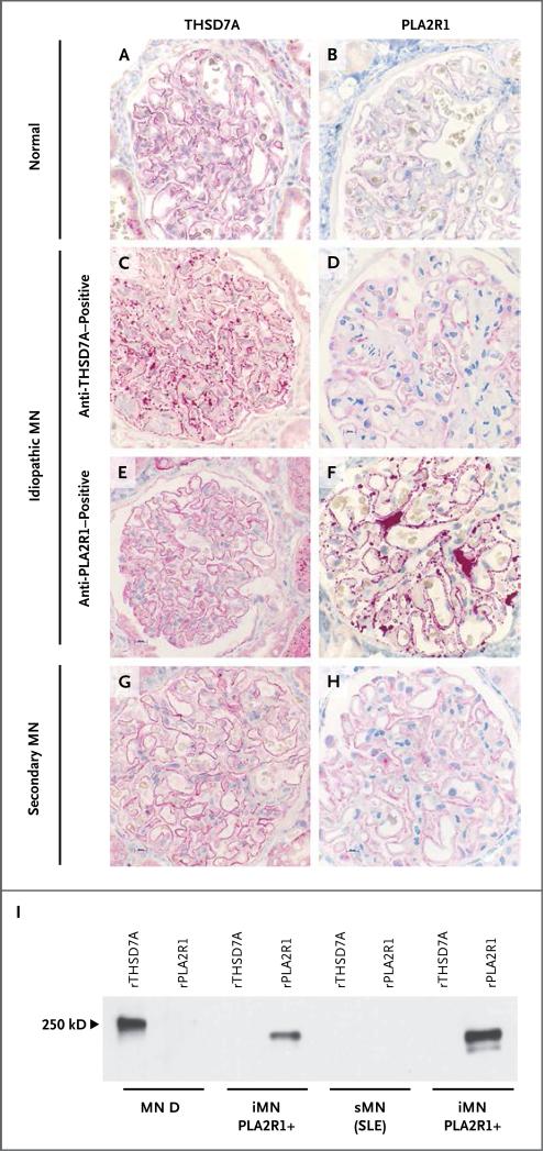
Figure 5. Expression Level of THSD7A and PLA2R1 in Patients with Idiopathic and Secondary Membranous Nephropathy and in a Healthy Control.
Immunohistochemical staining for THSD7A and PLA2R1 was performed in renal-biopsy specimens from healthy controls and from patients with membranous nephropathy. Panels A and B show normal staining for THSD7A and PLA2R1, respectively, in a control biopsy specimen. Panel C shows markedly enhanced staining for THSD7A in a patient with serum anti-THSD7A antibodies, whereas PLA2R1 staining, as shown in Panel D, is normal. Conversely, the biopsy specimen from a patient with serum anti-PLA2R1 antibodies shows that staining for THSD7A was not increased, as shown in Panel E, but staining for PLA2R1 was increased, as shown in Panel F. The biopsy specimen from a patient with secondary membranous nephropathy (sMN) due to systemic lupus erythematosus (SLE) and no detectable serum antibodies against THSD7A or PLA2R1 has normal staining for both antigens, as seen in Panels G and H. Panel I shows the reactivity of IgG eluted from patient biopsy samples with recombinant THSD7A and PLA2R1. Only the IgG eluted from the biopsy sample from a patient with idiopathic membranous nephropathy (iMN) and detectable serum anti-THSD7A antibodies recognized THSD7A (MN D; see also Table S3 in the Supplementary Appendix). The eluates from two biopsy samples from patients with idiopathic membranous nephropathy and serum anti-PLA2R1 autoantibodies recognized PLA2R1 but not THSD7A. IgG eluted from one patient with lupus membranous nephropathy and no detectable serum antibodies against THSD7A or PLA2R1 recognized neither antigen.

