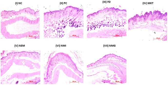
Figure 17. H&E histological staining showing altered keratinocyte proliferation and differentiation with IMQ exposure.
C57/BL mice were exposed to the IMQ suspension for 5 days followed by treatment. i) Negative control (NC) ii) Positive Control (PC) iii) Free Drug (FD) iv) Marketed gel (MKT) v) Nano emulsion (NEM) vi) Nano micelle (NMI) vii) Nano miemgel (NMG). Data represent mean±SD, n = 6, significant where *p<0.05.

