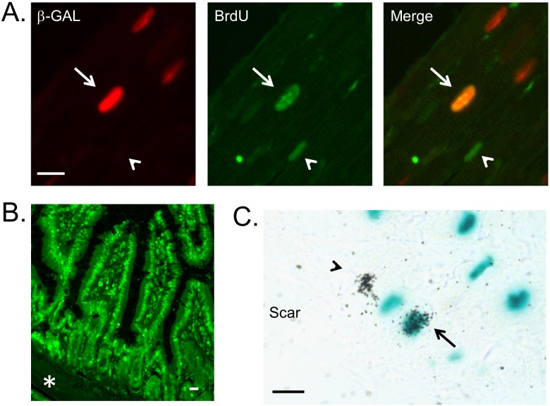
Figure 1. Examples of cardiomyocyte DNA synthesis assay.
(A) Use of BrdU to monitor cardiomyocyte DNA synthesis in non-injured adult mice receiving 9 consecutive daily injections of NRG1β1 (BrdU was delivered using a mini-osmotic pump). Left panel shows anti-β-galactosidase immune reactivity, middle panel shows anti-BrdU immune reactivity, and right panel shows the merged image. Arrow indicates a BrdU positive cardiomyocyte nucleus, arrowhead indicates a BrdU positive non-cardiomyocyte nucleus. Bar = 10 microns. (B) BrdU incorporation in the nuclei of the small intestine microvilli epithelial cells of an NRG1β1-treated mouse. Note the absence of BrdU signal in the muscularis mucosae zone (asterisk). Bar = 10 microns. (C) Use of 3H-Thy to monitor cardiomyocyte DNA synthesis in non-injured adult mice receiving 9 consecutive daily injections of NRG1β1 (3H-Thy was delivered as a single bolus 1 hour after the last NRG1β1 treatment). Arrow indicates a 3H-Thy positive cardiomyocyte nucleus, arrowhead indicates a 3H-Thy positive non-cardiomyocyte nucleus. Bar = 10 microns.

