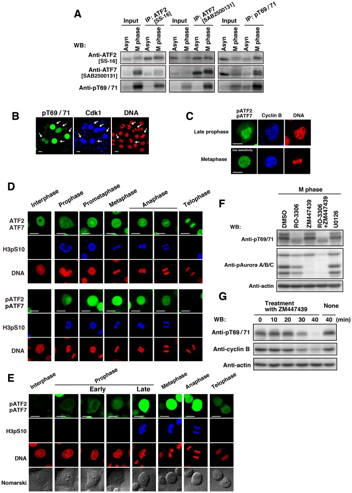
Figure 4. Phosphorylation of ATF2 and ATF7 from prophase to anaphase.
(A–E Cells were synchronized using single-thymidine block and released into thymidine-free medium for 11 h. (A) Mitotic cells collected by mitotic shake-off and asynchronous (Asyn) cells were lysed with Triton X-100. Endogenous ATF2, ATF7, and pATF2/pATF7 were individually immunoprecipitated from Triton X-100 cell lysates using antibodies specific for ATF2 [SS-16], ATF7 [SAB2500131], and pATF2/pATF7 (pT69/71). Full-length blots are presented in S11A Fig. The gels for ATF7 IP blotted with anti-pT69/71 antibody and anti-ATF2 and anti-ATF7 antibodies have been run under the same experimental conditions. (B) Cells were triply stained with anti-pT69/71 and anti-Cdk1 antibodies and PI (for DNA). Anti-Cdk1-stained cells are pseudo-colored as blue. Scale bars, 20 µm. Arrows indicate mitotic cells. (C) Cells were triply stained with anti-pT69/71 and anti-cyclin B1 antibodies and PI (for DNA). Staining of pATF2/pATF7 was recorded with a low sensitivity. Anti-cyclin B1-stained cells are pseudo-colored as blue. Scale bars, 20 µm. (D) Cells were triply stained with anti-ATF2[N96] (upper panels) or anti-pT69/71 (lower panels) antibody, anti-histone H3pS10 antibody (for M phase) and PI (for DNA). Scale bars, 20 µm. (E) Cells were triply stained with anti-pT69/71 antibody, anti-histone H3pS10 antibody (for M phase), and PI (for DNA). Scale bars, 20 µm. (F) Cells were arrested at G2 phase using 9 µM RO-3306 and were released into RO-3306-free medium containing 10 µM MG132. At 20 min after release, cells were treated for an additional 60 min in the presence of 10 µM MG132 together with DMSO, 9 µM RO-3306, 10 µM ZM447439, RO-3306 plus ZM447439, or 20 µM U0126. Whole cell lysates were analyzed by WB. Full-length blots are presented in S11B Fig. (G) Cells arrested at G2 phase by treatment with 9 µM RO-3306 were released into RO-3306-free medium containing 100 µM monastrol for 1 h. The monastrol-arrested cells were collected by mitotic shake off and incubated with 10 µM ZM447439 for the indicated times (induction of mitotic slippage). Whole cell lysates were analyzed by WB. Full-length blots are presented in S11C Fig.

