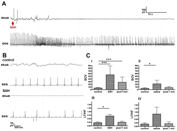
Figure 6. Cardiac responses to SAH induction.
Fig. 6 depicts a sample ECG trace after SAH. Three out of seven mice in the SAH group displayed transient high grade second and/or third degree atrioventricular block during flat-lining of the ECoG shortly after SAH (Figs. A and B bottom). F-test for comparison of equality of variances revealed increased SCV in SAH mice (C I) and in saline-injected mice (C II). SAH mice also display a transient significant increase in LF/HF ratio, potentially reflecting sympathetic activation to stabilize CPP after SAH or a Cushing reflex due to elevated ICP (C III).

