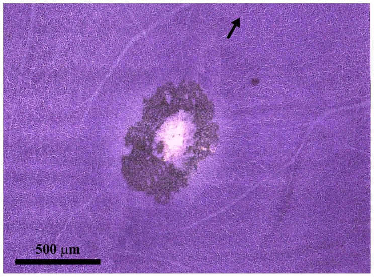
Figure 1. Foveal region of an Alouatta retina.
Retinal flat mount AC 02 Left Male stained by the method of Nissl using cresyl violet as stain. Some retinal pigment remained attached to the region surrounding the foveal pit. The arrow points towards the location of the optic nerve head, which is out of the field. The retinal raphé is located on the opposite side in relation to the fovea and is indicated by the convergence of retinal vessels.

