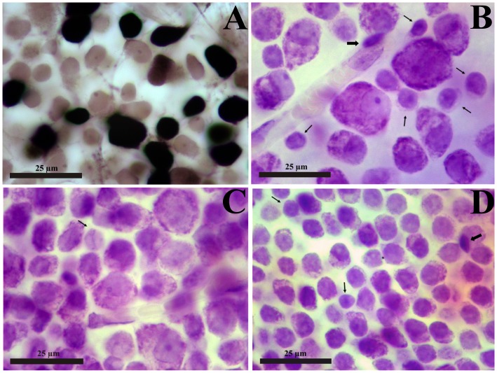
Figure 2. Retinal ganglion cell layer of New World monkeys.
(A) Retinal flat mount of a Cebus monkey (right retina, male animal), focus on the ganglion cell layer, 1.8 mm temporal to the fovea. Cells were labeled by Biocytin retrograde transport after deposits of the neurotracer in the optic nerve, 2 mm behind the eyeball, followed by 24 hs survival time [40]. The retina was dissected free from the other retinal layers and the retinal pigment epithelium, incubated in Vectastain ABC System solution, reacted for peroxidase histochemistry using diaminobenzidine as chromophore, and exposed to osmium tetroxide to intensify the reaction product. All retrograde labeled cells are retinal ganglion cells showed in the picture as black stained cell bodies (axons and dendrites are also labeled but are largely out of focus in this photomicrograph). Cells that were not retrograde labeled are the light brown cell bodies in the picture and were stained by the weak unspecific diaminobenzidine reaction intensified by the osmium tetroxide exposure: they comprised some ganglion cells, displaced amacrine cells, microglia, astrocytes, and endothelial cells. This procedure was used to consolidate the criteria for identify and counting ganglion cells and displaced amacrine cells in New World monkeys (see text for details). (B–D) Alouatta retinal flat mount (AC 04 Left Male), stained by the method of Nissl using cresyl violet as stain. The retinal ganglion cell layer is shown at three increasing eccentricities: 5 mm (B), 2 mm (C), and 1 mm (D) temporal to the fovea. Ganglion cells comprise a heterogeneous population of neurons readily distinguishable from other neurons (thin arrows), glial cells (thick arrows), and endothelial cells. The non-ganglion cell neurons of the retinal ganglion cell layer comprise displaced amacrine cells and displaced horizontal cells, the later relatively rare in comparison with the former [42]–[44]. The glial cells comprise microglia (thick arrow in B) and astrocytes (thick arrow in D), which are generally located internally (microglia) or externally (astrocytes and microglia) to the retinal ganglion cells. The endothelial cells can be distinguished by forming the blood vessel walls (B and C). In despite the large increase in ganglion cell density and decrease in the sizes of ganglion cell bodies, the criteria to distinguish between cell classes of the retinal ganglion cell layer can be applied throughout the retinal flat mount, including regions close to the fovea. In the foveal slope (D) the ganglion cells are stacked in 4–7 cell layers and careful focus through the ganglion cell layer thickness is necessary to perform precise neuronal counting.

