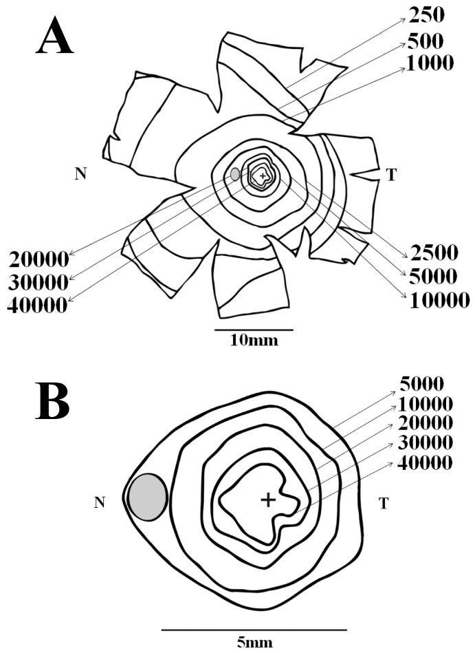
Figure 5. Ganglion cell isodensity maps for another Alouatta retina.
(A) Isodensity contours for the retina AC 02 Left Male. (B) Isodensity contours for the central retinal region. Conventions were the same of Fig. 4. Similarly to other retinas studied in this work, ganglion cell isodensity contours were slightly elongated in the nasal direction. In addition, in this retina there was also an elongation of the central isodensity contours along the dorsoventral meridian.

