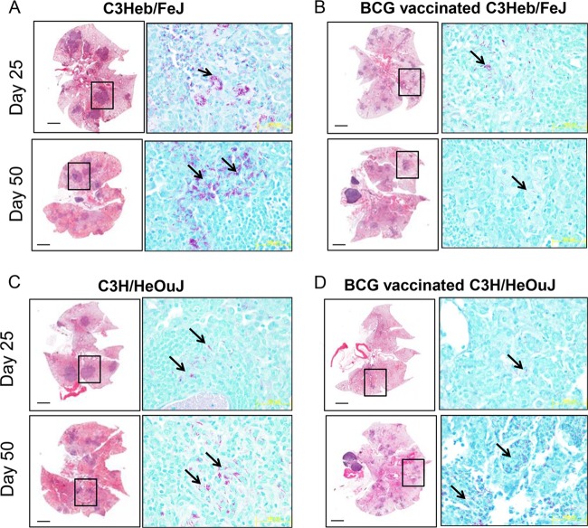
FIG 2.
Changes in lung pathology in control and BCG-vaccinated C3H/HeOuJ and C3Heb/FeJ mice. Shown are representative photomicrographs of hematoxylin-eosin-stained slides (left) and acid-fast staining of slides (right) from the lungs of control or vaccinated mice. (A) As early as 25 days after infection, significant areas of necrosis were observed in C3Heb/FeJ mice. As disease progressed (day 50), areas of necrosis and bacterial burden (denoted by acid-fast staining) significantly increased in C3Heb/FeJ mice. Clusters of bacilli can be observed, which accumulated in areas of necrosis (arrows). (B) In contrast, BCG vaccination of C3Heb/FeJ mice significantly diminished necrosis and bacillary loads on both days 25 and 50 after infection. (D) BCG vaccination limited lesion size and the presence of acid-fast bacilli in C3H/HeOuJ mice temporarily 25 days after infection, and this protection was lost during chronic infection. (C) In control C3H/HeOuJ mice, bacillary numbers increased at day 50 albeit to a much lesser extent than in C3Heb/FeJ mice. Magnifications, ×4 (left) and ×100 (right).

