FIG 2.
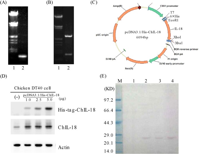
Construction and expression of a full-length ChIL-18 expression plasmid. (A) Lane 1, marker λDNA/EcoRI+HindIII; lane 2, PCR products of the full-length ChIL-18 gene. (B) Lane 1, marker λDNA/EcoRI+HindIII; lane 2, identification of pcDNA3.1/His-ChIL-18 recombinant plasmid digested with EcoRI and XhoI. (C) Structure of the pcDHA3.1/His-ChIL-18 recombinant plasmid. (D) Western blots were prepared with extracts from DT40 cells with untransfected (−) or transfected recombinant pcDNA3.1/His-ChIL-18 plasmid with different doses (1.0, 2.5, and 5.0 μg) for 48 h. The blots were then probed with antibodies against His tag, ChIL-18, and actin. (E) Western blot analysis of purified His-tagged ChIL-18 protein expressed in DT40 cells. Lane M, protein size marker; lane 1, empty expression plasmid pcDNA3.1/His, negative control; lanes 2 to 4, purified His-tagged ChIL-18 protein collection fractions by Ni-affinity chromatography.

