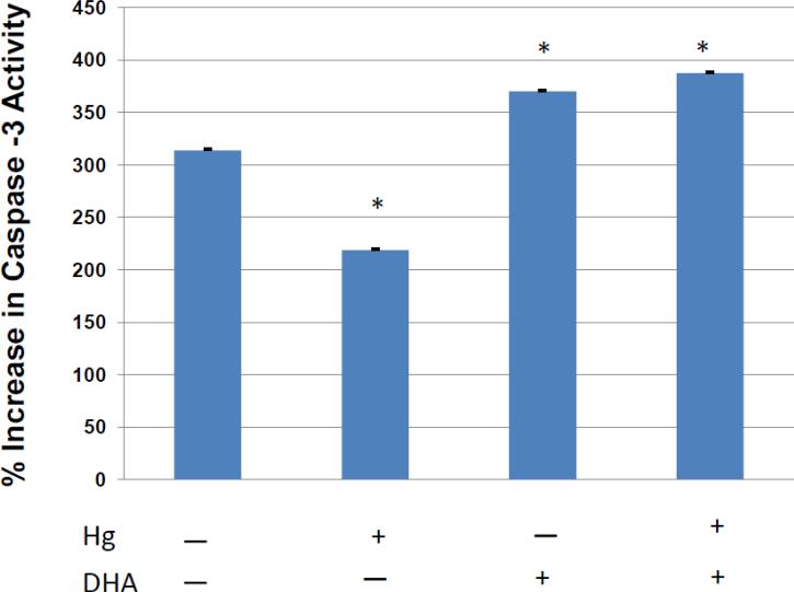
Figure 3.
Quantitative assessment of the effect of DHA on CD95 mediated caspase 3 activation in mercury burdened T cells. Jurkat T cells were incubated overnight with or without DHA. HgCl2 was then added to half the cells, and then cells were treated with anti-CD95 in order to stimulate CD95 signaling. After an additional culture period cells were assayed for active caspase 3, and the % increase in caspase 3 activity determined by comparison with control cells which were not stimulated with anti-CD95. Error bars represent the SEM of 6 replicate samples. The (*) symbol represents a statistically significant difference of the mean compared to the mean of Hg (−) and DHA (−) cells.

