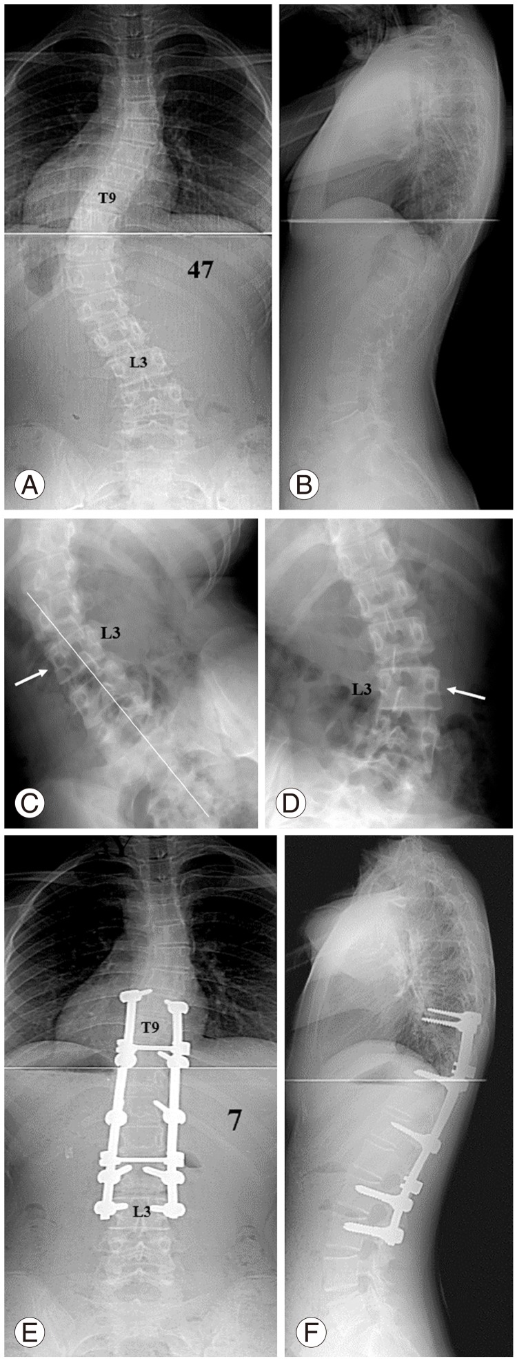
Fig. 1.
(A, B) Preoperative standing anteroposterior and lateral radiographs of a 13.3-year-old girl with 47° thoracolumbar curve. (C) L3 (arrow) crossed the mid-sacral line in the right bending radiograph. (D) L3 (arrow) rotated less than Nash-Moe grade II, and it actually rotated in the opposite direction in the left bending radiograph. (E, F) Anteroposterior and lateral radiographs taken at 3.5 years after operation. The fusion was extended down to L3. The thoracolumbar curve was corrected to 7° with well-balanced spine and horizontalization of the lowest instrumented vertebra.

