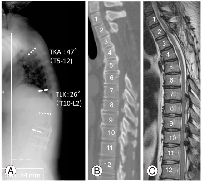
Fig. 2.
(A) Radiograph of the spine showing 47° thoracic kyphosis (TKA, T1-12), 26° thoracolumbar kyphosis (TLK, T10-L2) and 84 mm of sagittal vertical axis (SVA). (B, C) Computed tomography and magnetic resonance imaging showing ossification of the posterior longitudinal ligament from C2 to C5 and T3 to T6. The spinal cord was compressed by the ossification of the ligamentum flavum at T1-3 and T7-11.

