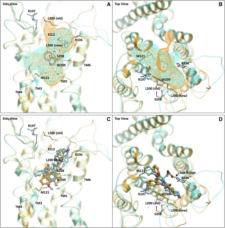
Fig. 7.
Molecular model of T-0632 bound to CCK1R. In all images, the new model described in this work (Supplemental Fig. 1) is represented in cyan ribbon and stick and the old model (Supplemental Fig. 2) (Cawston et al., 2012) is displayed in gold ribbon and stick. The viewpoints are from the TM “side” between TM4 and TM5 (A and C) and from the “top” through the N-terminal extracellular surface (B and D). (A) and (B) illustrate the comparison of the allosteric binding pockets of the old (gold wire mesh) and new (cyan wire mesh) models. A change in volume and shape was observed due to the movement of ECL2. (C) and (D) illustrate the comparison of the binding poses of benzodiazepine (gold stick) and T-0632 (cyan stick) in the old (gold ribbon) and new (cyan) models, respectively. A salt bridge was shown to be present between the carboxylate of T-0632 and Arg336 of CCK1R.

