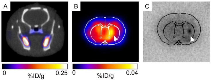
Figure 9.
(A) Very low brain uptake is observed in 52Mn-based PET/CT imaging in vivo. (B) When the brain is excised and imaged ex vivo, low levels of brain uptake are detectable. Both ex vivo imaging and autoradiography indicated that increased levels of 52Mn are retained in the vicinity of hNPC-DMT1 (white arrowhead, B and C).

