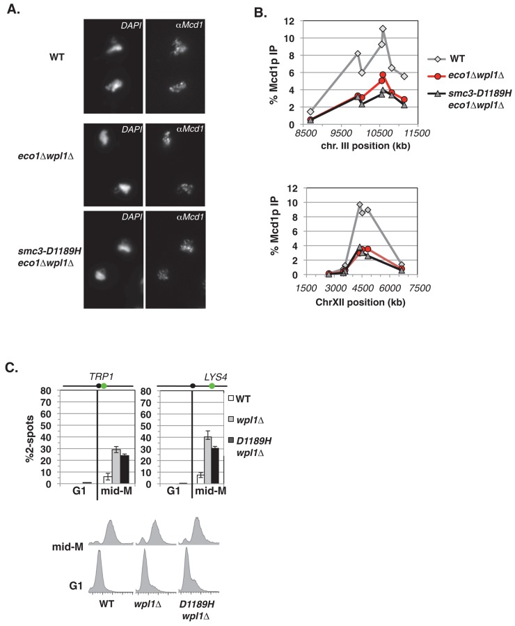
FIGURE 5:
Smc3-D1189H fails to suppress the characteristic wpl1∆ defects of reduced cohesin binding and partial cohesion loss. (A–C) Haploid wild-type (WT; VG3349-1B), eco1∆ wpl1∆ (VG3503-4A), and smc3-D1189H eco1∆ wpl1∆ (VG3549-7A) cells were arrested in mid–M phase as described in Figure 1C. (A) Chromosome spreads of mid–M phase cells. WT (top), eco1∆ wpl1∆ (middle), and smc3-D1189H eco1∆ wpl1∆ (bottom). Cells were processed to detect chromosomal DNA (4′,6-diamidino-2-phenylindole) and cohesin using αMcd1p antibodies. (B) ChIP of Mcd1p in mid–M phase cells. Cells were fixed and processed for ChIP using αMcd1p antibodies. WT (gray line, gray diamonds), eco1∆ wpl1∆ (red line, red circles), and smc3-D1189H eco1∆ wpl1∆ (black line, gray triangles). Mcd1p binding was assessed as described in Figure 4C. Top, chromosome III pericentric CARC1. Bottom, chromosome XII CEN-distal CARL. (C) Effect of smc3-D1189H on cohesion loss in a wpl1∆ background. Haploid WT, wpl1∆, and smc3-D1189H wpl1∆ cells were arrested in mid–M phase as described in Figure 1C. Left, cohesion loss at CEN-proximal TRP1 locus assayed in haploid WT (VG3460-2A), wpl1∆ (VG3604-4C), and smc3-D1189H wpl1∆ (VG3605-5D) strains. Right, cohesion loss at CEN-distal LYS4 locus assessed in haploid WT (VG3349-1B), wpl1∆ (VG3626-2E), and smc3-D1189H wpl1∆ (VG3627-3C) strains. Bottom, DNA content. The percentage of cells with two GFP signals (sister separation) is plotted. Data were derived from two independent experiments; 100–300 cells were scored for each data point in each experiment.

