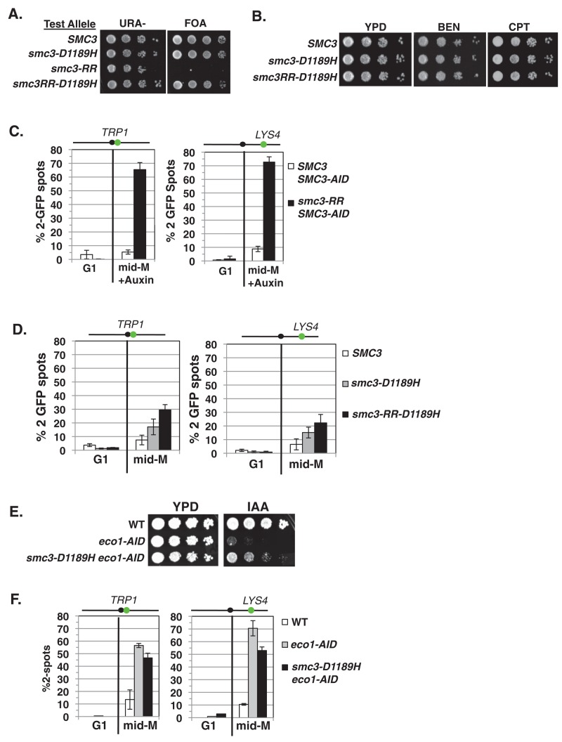
FIGURE 6:
smc3-D1189H robustly suppresses the cohesion defect of the smc3-K112R, K113R (RR) mutation. (A) Plasmid shuffle to assess viability of the chimeric smc3-RR-D1189H allele. Haploid shuffle strain VG3464-16C bearing plasmid pEU42 (SMC3 CEN URA3) and a second SMC3 “test allele,” SMC3, smc3-D1189H, smc3-RR, or chimeric smc3RR-D1189H, was grown and plated as described in Figure 2A onto URA–dropout or FOA-containing media. Plates were incubated 3 d at 23°C. (B) Assessment of drug sensitivity. Haploid SMC3 (MB45-1A), smc3-D1189H (MB46-1A), or chimeric smc3-RR-D1189H (MB47-1A) strains grown and plated as described in Figure 2A onto YPD, BEN, and CPT and incubated at 30°C for 2 d. (C). Cohesion loss of in smc3-RR cells. Haploids bearing SMC3-AID and a second SMC3 allele, either WT or smc3-RR, were depleted for SMC3-AID from G1 through mid–M phase arrest. Left, cohesion loss at CEN-proximal TRP1 locus assessed in haploid SMC3 SMC3-AID (MB84-1A) and smc3-RR SMC3-AID (MB83-1A) strains. Right, cohesion loss at CEN-distal LYS4 locus assessed in haploid SMC3 SMC3-AID (MB81-1A) and smc3-RR SMC3-AID (MB79-1A) strains. The percentage of cells with two GFP spots (sister separation) is plotted. (D) Cohesion loss of smc3-RR-D1189H in mid–M phase–arrested cells. Haploid SMC3, smc3-D1189H and chimeric smc3-RR-D1189H were arrested in mid–M phase as described in Figure 1B. Left, cohesion loss at CEN-proximal TRP1 locus assessed in haploid wild-type (SMC3; MB65-1A), smc3-D1189H (MB66-1A), or chimeric smc3-RR-D1189H (MB67-1A) strain. Right, cohesion loss at CEN-distal LYS4 locus assessed in haploid strains from B. The percentage of cells with two GFP spots (sister separation) is plotted. (E, F) Assessing smc3-D1189H ability to suppress Eco1p depletion. (E) Viability after Eco1p depletion. Haploid WT (VG3620-4C), ECO1-AID (VG3662-1D), and smc3-D1189H ECO1-AID (VG3663-2E) were grown and plated as described in Figure 2A onto YPD alone or containing auxin (750 μM) and then incubated at 23°C for 2 d. (F) Cohesion loss after auxin-mediated Eco1p depletion from G1 cells through mid–M phase arrest. Left, cohesion loss at CEN-proximal TRP1 locus assayed in haploid strains WT (VG3460-2A), ECO1-AID2 (VG3659-1A) and smc3-D1189H ECO1-AID2 (VG3663-2E) strains. Right, cohesion loss at CEN-distal LYS4 locus assessed in haploid WT (VG3620-4C), ECO1-AID2 (VG3646-1A) and smc3-D1189H ECO1-AID2 (VG3650-1E) strains. The percentage of cells with two GFP signals (sister separation) is plotted. (C, D, F) Data were derived from two independent experiments; 100–300 cells were scored for each data point in each experiment.

