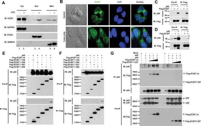
FIGURE 1:
ECSIT interacts with p65/p50 NF-κB proteins after LPS stimulation. (A) HEK293-TLR4 cells were treated or not with 100 ng/ml LPS for 45 min, and then cytosol (Cyt), nuclear (Nuc), and mitochondrial (Mito) fractions were isolated, followed by immunoblot (IB) with antibodies to ECSIT, GAPDH, PCNA, and GRIM19. (B) HEK293-TLR4 cells were treated or not with100 ng/ml LPS for 45 min, and a confocal microscopy analysis was performed. (C) HEK293T cells were transiently transfected with Flag-ECSIT or p65-expression vector, as indicated, and then the immunoprecipitation (IP) assay was performed with anti-Flag antibodies, followed by IB with antibodies to anti-p65 or anti-Flag. (D) HEK293T cells were transiently transfected with Flag-ECSIT or p50-expressing vector, as indicated, and the IP assay was performed with anti-Flag antibodies, followed by IB with antibody to anti-p50 or anti-Flag. (E) HEK293T cells were transiently transfected with Flag-ECSIT wt or Flag-ECSIT truncated mutants, along with the p65-expressing vector, as indicated, and the IP assay was performed with anti-Flag antibodies, followed by IB with antibody to anti-Flag or anti-p65. (F) HEK293T cells were transiently transfected with Flag-ECSIT wt or Flag-ECSIT truncated mutants, along with p50-expressing vector, as indicated, and the IP assay was performed with anti-Flag antibodies, followed by IB with antibody to anti-Flag or anti-p50. (G) HEK293T cells were transiently transfected with p65-expressing vector, p50-expressiong vector, different concentrations of Flag-ECSIT wt, or different concentrations Flag-ECSIT 1-257, as indicated, and the IP assay was performed with anti-p65 antibody, followed by IB with antibodies to anti-Flag, anti-p50, and anti-p65.

