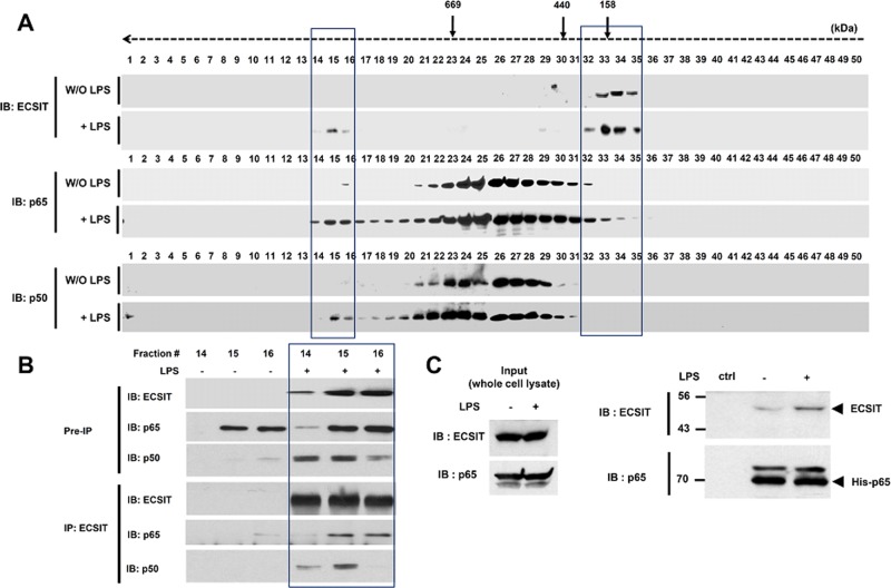
FIGURE 2:
ECSIT endogenously forms the ECSIT/p65/p50 complex and interacts with the p65/p50 NF-κB proteins. (A) THP1 cells were treated for 60 min or not with LPS. The cell extracts were prepared and fractionated through a Superose6 10/300 GL column. Each fraction (40 μl) was analyzed by immunoblot (IB) with the indicated antibody to anti-ECSIT, anti-p65, or anti-p50. Apparent molecular mass was evaluated after column calibration with standard proteins: thyroglobulin (669 kDa), ferritin (400 kDa), catalase (232 kDa), and aldolase (158 kDa). The elution positions of these proteins are indicated at the top. (B) Endogenous immunoprecipitation (IP) of ECSIT from fractions 14–16 prepared from THP-1 cells, as described in A, followed by IB with antibodies to anti-ECSIT, anti-p65, or anti-p50. (C) The His-tagged p65 pull-down assay was performed with whole-cell lysates prepared from THP-1 cells treated or not with LPS (100 ng/ml) for 45 min, and then IB with antibody to anti-ECSIT or anti-p65 was performed.

