Figure 2.

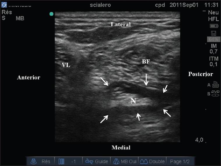
Ultrasonic typical doughnut image. Hypo-echoic 20 mL local anesthetic typically surrounded the sciatic nerve at mid-thigh (arrows). VL: Vastus lateralis; BF: Biceps femoris; N: Nerve

Ultrasonic typical doughnut image. Hypo-echoic 20 mL local anesthetic typically surrounded the sciatic nerve at mid-thigh (arrows). VL: Vastus lateralis; BF: Biceps femoris; N: Nerve